What Is the Inclination Joint of a Microscope?
The inclination joint of a microscope is part of the microscope’s base. Not all microscopes have this part, but many do, especially compound microscopes. The inclination joint is instantly recognizable, as it is a pin that extends from the base of the microscope. Read on to learn more on how to operate the inclination joint, the history of the microscope, and other microscope parts.
How Do You Operate an Inclination Joint?
The inclination joint is a pin that sticks out toward the top of the base. If you wish to tilt your microscope to get a different angle of what you’re viewing, put one hand on the base to keep it steady. With the other hand, pull the pin out of its slot and tilt the microscope back. Be careful, however—using an inclination joint and pulling it back too far often results in the samples falling off the slide. If you intend to pull it back very far, use a small piece of tape on the slide itself to keep samples in place.
What Are the Other Parts of the Base?
In addition to the inclination joint, microscopes have other parts that are considered part of the base. The base itself is designed in such a way as to steady the microscope for more stable viewing. Attached to it is the pillar, which is the vertical part that extends from the base. Many compound microscopes also have a mirror on the base, although it can be on the end of the arm of the microscope. Its job is to reflect light into a microscope for viewing. Each base also has two feet to help keep it steady.
What Are More Key Microscope Terms?
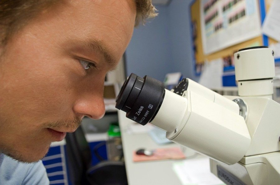
In order to operate a microscope correctly, you’ll need to know a few more key parts of the microscope. The top of the microscope, or the piece that you look through, is called the eyepiece. This connects to the body and tubes. The arm of the microscope is the piece that comes out of the pillar, and it forms somewhat of a U shape. There are two knobs on the arm, both the fine and coarse adjustment knobs, which is what you use to bring slides into focus. The place where you put your sample (or slide) is called the stage, and there are two clips on either side of it to keep samples in place. The nose piece is the part of the microscope that you turn (immediately above the sample), and it has two lenses, a low-power and high-power objective.
How Do You Operate a Microscope?
To operate an everyday compound microscope, you would first look through the eyepiece and adjust the mirror to make sure there’s enough light in the microscope to view samples. You also want to make sure there’s no dust where you’ll be putting the sample, like the stage or clips. Next, move the nose piece to ensure the low-power objective in line with the sample. To bring the slide into focus, first adjust the coarse adjustment knob, followed by the fine adjustment knob.
What Is the History of the Microscope?
The microscope was first invented in the late 16th century by Hans Lippershey, who was a spectacle maker, so that he could magnify images. It’s true that a magnifying glass is a bit of rudimentary microscope, as it also magnifies something you’d wish to view, but a higher level of magnification was needed in order to study objects more closely. Over the years, many different types of microscopes have been invented.
What Are Different Types of Microscopes?
In botany or in a classroom setting, the compound type of microscope is the most common. The first type of microscope is a simple microscope, which is a magnifying, convex lens combined with a place to hold specimens or samples. A compound microscope builds upon this version by adding a second lens to magnify the image of the first. Another type of microscope is a stereo microscope, which has 300x magnification for larger specimens. Other types include confocal microscopes, which use laser light, scanning electron microscopes, which use electrons, and transmission electron microscopes, which also use electrons along with vacuum-scanned specimens for better transparency.


