What Is Liver Hypodensity?

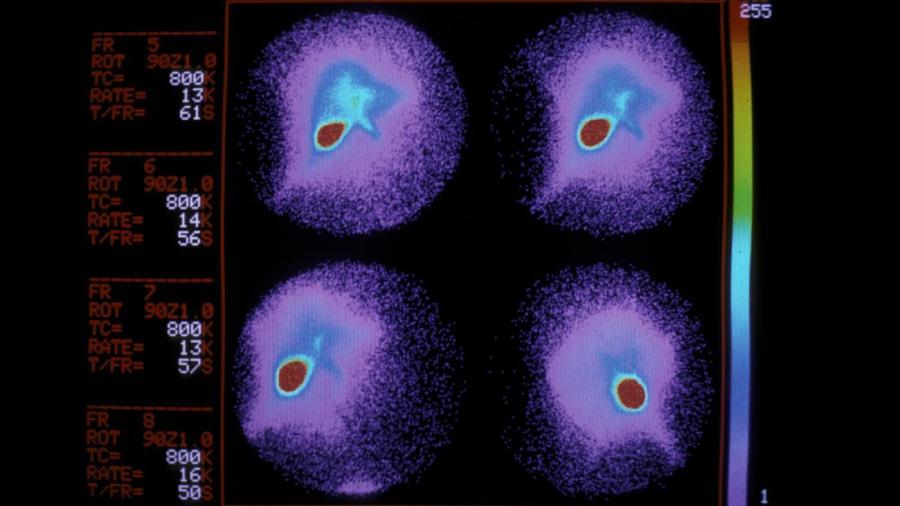
Liver hypodensity is a term used by radiologists to describe areas of the liver on CT scans, according to Dr. Tracy A. Berg. It means there are areas of the liver that appear less dense on a CT scan than the surrounding liver tissue.
Liver hypodensity on a CT scan is caused by a variety of factors, according to Radiopaedia, such as benign and malignant neoplasms, focal nodular hyperplasia, focal fatty infiltration, benign epithelial cysts, cysts caused by liver disease and infection, pyogenic abscesses, fungal abscesses, amoebic abscesses, vascular infarctions, lacerations, old hematomas, and biliary tree dilation. Liver hypodensity is usually caused by the presence of hepatic fat, according to Radiopaedia. Dr. Jeffrey Fine indicates that liver hypodensity is also caused by benign cysts.
CT machines use X-rays to circle the body to create image slices that are then stacked on each other to create a three-dimensional image. They provide greater detail of internal organs than traditional X-rays and are more useful in diagnosing injuries and diseases of the liver, according to Johns Hopkins Medicine. They can also be used to guide the placement of needles in the liver for biopsies, the withdrawal of fluid or to diagnose the causes of jaundice.


