How Is a Bone Density Test Done?
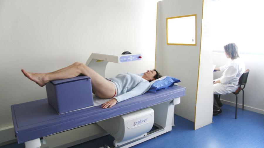
With a central bone density scan, the patient lies on a table while the scanner passes over his body, emitting X-rays to test the density of bones in the lower spine and hips, according to MedlinePlus. The purpose of the test is to detect osteoporosis and predict fracture risks.
Bone density tests are painless, fast and easy, according to Mayo Clinic. They require little-to-no preparation by the patient; however, the patient should inform his doctor if he recently had imaging tests with barium or contrast media, as these materials affect the outcome of the bone density test. If the doctor orders a peripheral bone density test, the test uses a small portable device.
While standard X-rays eventually show bone loss, the bone density test detects weakened bones earlier when treatment is still an option, according to Johns Hopkins Medicine Health Library. Once the test is complete, a computer calculates the bone density based on the number of photons absorbed by the bones. The computer compares the results with those of the average young adult of the same sex and ethnic background. The doctor sometimes orders other tests, including blood tests, at the same time as the bone density test. These tests determine contributing factors to bone loss.


