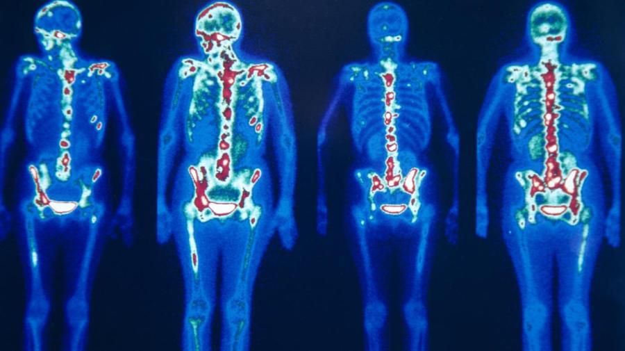
What Is a Nuclear Bone Scan?

A bone scan is a nuclear imaging test used in the diagnosis of bone diseases and disorders. A bone scan is also used in tracking the location of cancer that has metastasized, or spread to a different location than the original diagnosis, notes Mayo Clinic.
Determining the cause of unexplained bone pain with a bone scan allows examination of the entire skeleton. Bone disorders can include avascular necrosis, fractures, fibrous dysplasia, arthritis and Paget’s disease of bone, osteomyelitis. They can also include cancer of the bone and cancer metastasizing to the bone from another area, according to Mayo Clinic.
A bone scan poses no greater risk to the patient than a regular X-ray. The radiation exposure from the tracer used during the procedure is less than half of the radiation exposure experienced during a CT scan. Preparation for the procedure requires removing jewelry and metal objects and notifying the physician of pregnancy or if nursing an infant, explains Mayo Clinic.
The procedure starts with an injection of the tracer fluid containing small radioactive tracer into the veins. The tracers are then taken up in the procedure by cells and tissues, with the highest amount taken up by those repairing themselves. The time between the injection and the reading depends on the test ordered by the physician. Preliminary images are captured in some cases, but the main images are taken about two to four hours after injection, following circulation and absorption of the tracer into the bones, states Mayo Clinic.


